A study finds that T cells induced by heroin cross the blood-brain barrier to wreak havoc on the brain, hinting at new ways to prevent withdrawal.
ABOVE:© ISTOCK.COM, D-KEINE
Researchers have uncovered one way that opioid use seems to result in withdrawal symptoms, identifying a previously unknown pathway in the immune system that results in unstable and dysfunctional connections among brain cells.
Although the immune system has long been implicated in opioid withdrawal, the new findings, published January 19 in Cell, are the first to link the immune system’s interactions with the central nervous system and especially the blood-brain barrier to withdrawal, explains immunopathogenesis researcher Luis Montaner, who is a professor at the Wistar Institute in Philadelphia and didn’t work on the study. While some of the findings still need to be replicated and verified, Montaner says that the study presents scientists with “a roadmap for new clinical interventions to be tested” to prevent withdrawal and help people recovering from opioid addiction or dependence safely wean off of the drug and avoid reuse.
“The work represents a major advance in the emerging field of neural-immune interactions and the role of immune cells and mediators in modulating neural processes during opioid exposure,” Temple University immunologist and substance abuse researcher Toby Eisenstein, who also didn’t work on the study, tells The Scientist. “I think the paper is really a huge step forward.” Eisenstein says she was intrigued by the paper, which links withdrawal symptoms to an inflammatory immune response, because, as she wrote in a 2019 review article on the immune effects of morphine, decades of research have shown opioids and the immune cells implicated in the new study to be immunosuppressive, instead.
See “The Quest for Safer Opioid Drugs”
In the study, the researchers examined the blood of 21 human heroin users and compared it to that of 20 controls, finding that the former group’s blood contained various biomarkers suggesting their immune systems were in disarray (As of this article’s publication, neither of the paper’s corresponding authors have responded to The Scientist’s request for an interview). Investigating further with a series of flow cytometry experiments, they found that the heroin-using group’s blood contained high levels of an unusual immune cell called fragile-like regulatory T cells (Tregs). Tregs are usually involved in immunosuppression, but ones in this fragile-like state—which had previously only been observed in tumor microenvironments—lose their suppressive functions and instead produce the inflammatory cytokine interferon-γ (IFN-γ). The study authors write that opioid-induced hypoxia may have caused the cells’ unexpected state, as hypoxia has been implicated in triggering fragile-like states in the Tregs near tumors.
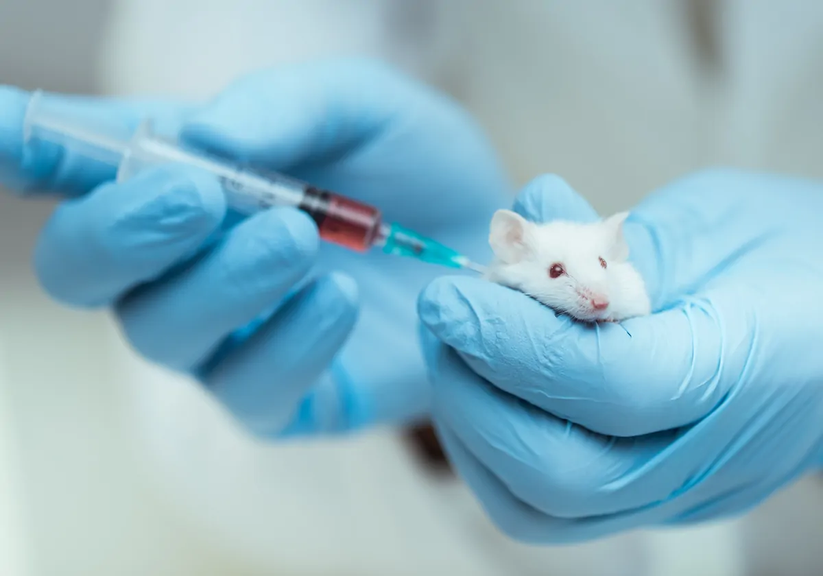
Moving over to a mouse model, the researchers found that treating mice with heroin resulted in higher fragile-like Treg counts and, therefore, higher IFN-γ expression in the bloodstream. Analysis of samples taken from the heroin-treated mice revealed that IFN-γ was not only more prevalent in the bloodstream, but also in the nucleus accumbens, a brain region that modulates goal-directed behaviors and reward pathways, making it relevant to understanding and treating addiction.
According to the study authors, the elevated IFN-γ in that region indicates that the fragile-like Treg cells were able to cross the blood-brain barrier, the protective layer that physically protects the brain from pathogens (and many pharmaceuticals) traveling through the body’s vasculature. The study linked the vulnerability to openings in the blood-brain barrier caused by C-C motif chemokine ligand 2 (Ccl2) expression by nucleus accumbens neurons, which itself was facilitated by opioid exposure, and which increases Treg trafficking into the brain. That stood out as particularly interesting to Eisenstein, she says: Chemokines are responsible for trafficking immune cell subsets that express the receptor for the respective chemokine, but the fact that the chemokine expression was coming from within the brain hints that brain cells have a larger role in the immune response than previously assumed.
“I think the paper is really advancing the whole neuroimmune field and maybe our understanding of what immune mediators and cells are doing in the brain,” Eisenstein says.
Staining revealed that levels of other cytokines and inflammatory agents were unchanged, which the researchers took to mean that the fragile-like Treg cells (and specifically the IFN-γ they expressed) were driving changes in the nucleus accumbens—weakening synaptic connections among neurons—that then resulted in withdrawal symptoms in the mice. In short, as the paper puts it, “Opioid stimulation . . . boosts the entry of fragile-like Tregs into the [central nervous system] and thereby contributes to structural and behavioral changes.”
With the mechanism pieced together, the researchers also showed that they could break it down, at least in mice. In both IFN-γ knockout mice and mice treated with an IFN-γ-neutralizing antibody prior to opioid treatment, neural connects remained strong and unchanged throughout withdrawal even though the fragile-like Treg cells still infiltrated the brain. In these experiments, mice displayed reduced and shorter-lived withdrawal symptoms—in one case for just 12 hours instead of 60.
See “Opioid Vaccines as a Tool to Stem Overdose Deaths”
The central question that remains, Montaner says, is whether breaking any one link in this mechanistic chain can prevent withdrawal symptoms in people as well as it does in mice. In that regard, Montaner says he’d like to see longitudinal studies in humans to determine whether fragile-like Treg cell count predicts withdrawal severity and duration as the new study seems to suggest it might.
The paper notes that it’s not clear what actually drives the expansion of fragile-like Tregs in the bloodstream of heroin users—Montaner notes that the study took samples from people days after they stopped using heroin, meaning they’d already started to experience withdrawal, but not while they used an opioid—an omission that makes it harder to understand the beginning stages of the process. Therefore, linking the cells’ presence to hypoxia represents a bit of an extrapolation on the part of the study’s authors, he says, as do some of the assumptions made about the human cohorts’ withdrawal experiences.
I think the paper is really advancing the whole neuroimmune field and maybe our understanding of what immune mediators and cells are doing in the brain.
—Toby Eisenstein, Temple University
Eisenstein notes that there’s plenty of literature—she coauthored much of it—showing that opioid use and subsequent withdrawal can suppress the immune system to the point that harmful gut bacteria leak out into the body, resulting in the inflammatory condition sepsis. While the new study didn’t look at the role of bacteria or sepsis in withdrawal, Eisenstein suggests that the two could potentially be related and that, hypothetically speaking, sepsis earlier in the withdrawal process could feasibly kick off or contribute to the inflammatory response the study authors observed days later, when they started collecting data.
“I think their conclusion that they reached is valid,” Eisenstein says. “But maybe, there’s another part of the mechanism they haven’t thought of, which is actually backwards from where they are. They say these fragile T cells get induced, then move from the periphery to the nucleus accumbens, then [express] interferon. But maybe these fragile T cells are induced from the sepsis phenomenon ahead of it.”
Also, the causative findings in mice may not translate to or explain the correlation identified in human heroin users, argues Irma (Lisa) Cisneros, an addiction researcher who studies how central nervous system immune cells respond to toxins and viruses.
“At the physiological level, everything is connected,” Cisneros tells The Scientist over email. “For example, the gut brain axis, or how metabolites from drug use and misuse in the liver can cross the [blood-brain barrier]. This manuscript almost skims the surface of all those aspects, but the link is hard to grasp. Especially given that one of the most important factors, in my opinion, is the neurocircuitry of addiction, which is not highlighted in this manuscript. Individuals do not use a drug the first time and get addicted, it is a process and reshaping of the neurocircuitry that results in addiction. It is more likely than not that neuroinflammation influences this neurocircuitry.” She adds that the brain circuitry of mice may not fully resemble that of a human with addiction, since they were treated with heroin rather than becoming addicted to it on their own.
Still, unanswered questions and tentative hypotheses aside, the study still shows immense potential for withdrawal and addiction research, Montaner argues.
“They highlight a lot of potential therapeutic targets that could be pursued in the future to test the hypothesis ‘Would these interventions affect the cycle of withdrawal?’” he says. “If the answer to that question is ‘Yes,’ then the implication is pretty large because withdrawal symptoms are what lead people to reuse again.”
Montaner says that it would be fairly straightforward to check whether fragile-like Treg cell count predicts withdrawal severity, and that he plans to do so in his own upcoming research. Also, he adds, any team that still has samples from previous withdrawal studies could go back and look for the cells as well. “That type of question, we couldn’t ask before this [study],” he says.
Meanwhile, Eisenstein suggests that, based on the results of the new study, testing IFN-γ blocking drugs on human opioid or heroin users and seeing if it prevents or mitigates withdrawal would be a worthwhile endeavor.
Correction (1/26/23): This article has been updated to accurately reflect the role of chemokines.

